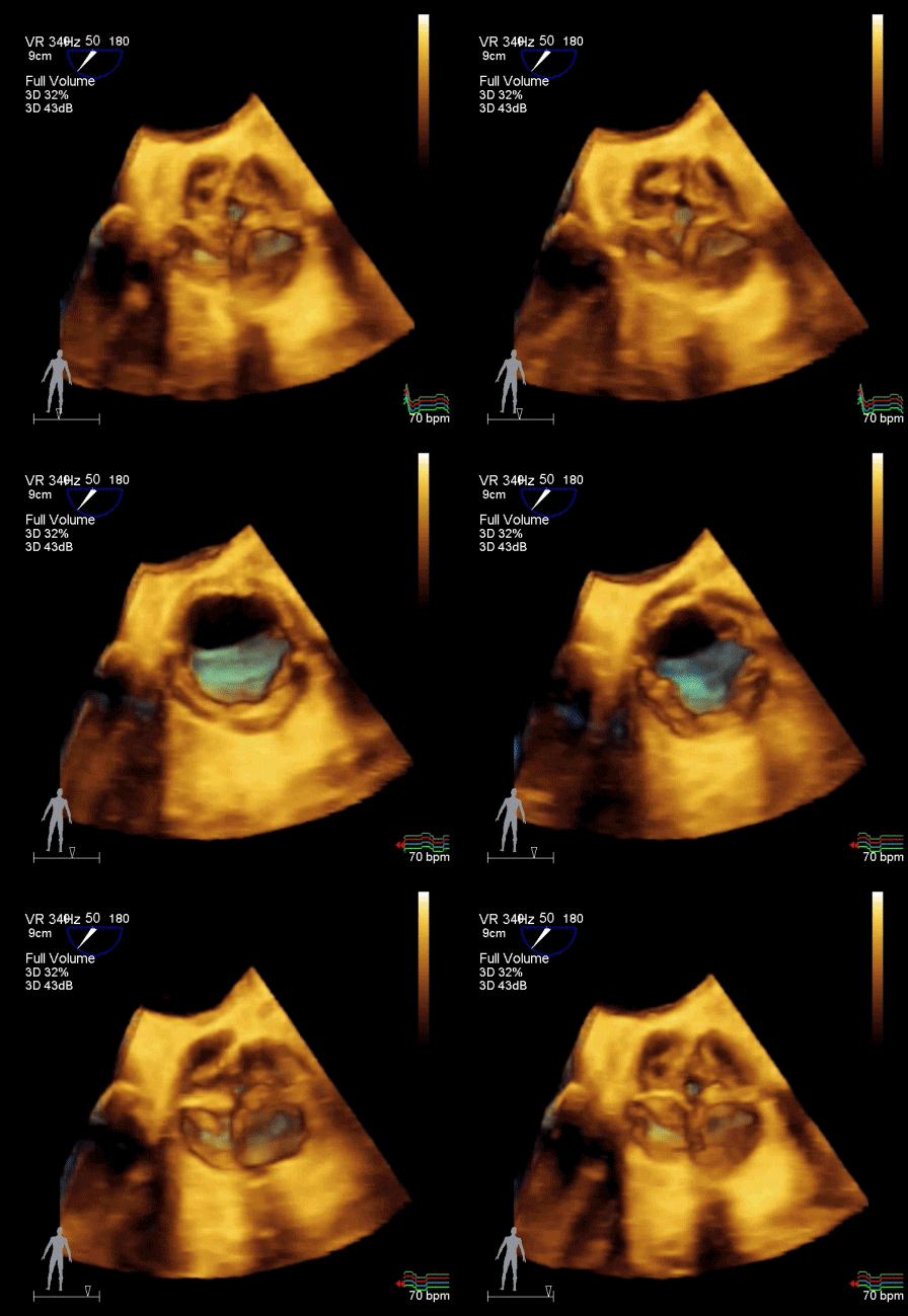
Echocardiogram
An echocardiogram (or “echo”) is an ultrasound of the heart. Some of the most common reasons for this test include shortness of breath, leg swelling, decreased stamina, heart murmur, and an abnormal EKG. An echocardiogram can assess known issues, such as heart failure or abnormal valves, or screen for problems in high risk individuals (e.g. those taking certain chemotherapies).
There are three main types of echocardiogram: a transthoracic echocardiogram, a stress echocardiogram, and a transesophageal echocardiogram.

What You Need to Know Before Your Echocardiogram
To ensure the best experience possible, please note the following important points:
If you're getting a stress echocardiogram, you'll need to skip certain medications.
If you’re scheduled for a stress echocardiogram please do not take any of the following medications on the morning of your test: metoprolol (Toprol), carvedilol (Coreg), atenolol (Tenormin), propranolol (Inderal), nebivolol (Bystolic), diltiazem (Cardizem), verapamil (Calan).
If you're getting a transesophageal echocardiogram, you'll need a ride home.
A transesophageal echocardiogram requires sedation. You’ll be groggy for an hour or two after your procedure, so you’ll need someone to drive you home.
Video Explainers
Echocardiogram: Getting The Test. Scheduled for an echocardiogram? This video will explain how the test works and what you can expect.
Echocardiogram: Understanding the Results. After your echocardiogram, Dr. Kelly will provide a complete report and interpretation. Although any important issues will be explained in detail, this video will explain how to understand the entire report.
Stress Echocardiogram. This video explains what to expect on the day of your stress echocardiogram, and how this test can help determine the likelihood that you have a blocked artery on your heart.
Transesophageal Echocardiogram (TEE). This video explains how this test works and what you should expect once you get to the hospital.
More Details
What is a transthoracic echocardiogram?
A transthoracic echocardiogram is performed by a sonographer (ultrasound imaging specialist). It is called transthoracic because the pictures are obtained by applying ultrasound through the chest wall, or thorax. This test assesses the structure and function of the heart’s chambers and valves. It can diagnose heart failure, heart enlargement, valve stenosis (stiffness), valve regurgitation (leakiness), and more. It can also assess the major blood vessels arising from the heart, such as the aorta and pulmonary artery.
You will remove your shirt and lie down on a stretcher. The sonographer will apply gel to the skin over your heart, then use an ultrasound probe to record both still pictures and videos of your heart in action.
You may be asked to roll from side to side, or to hold deep breaths, in order to get the best possible images. Don’t be surprised if you hear whooshing noises and beeps coming from the machine as the sonographer measures flow through different heart valves and saves pictures for the physician to review.
The test is completely safe, and there are no known risks. It generally takes 30-45 minutes to complete. You will receive your results within 2-3 business days.
What is a stress echocardiogram?
A stress echocardiogram is a transthoracic echocardiogram performed immediately after a stress test, while your heart is beating faster and harder than usual. We look for any parts of the heart that don’t seem to be functioning as well as the others, which can indicate inadequate blood flow secondary to a blocked coronary artery. Stress tests are described in more detail here.
What is a transesophageal echocardiogram?
A transesophageal echocardiogram (TEE) is an imaging test performed directly by Dr. Kelly. It is called transesophageal because the pictures are obtained by inserting an ultrasound probe down the throat and into the esophagus. From there, ultrasound waves are used to visualize the heart in motion.
Like a transthoracic echocardiogram, a transesophageal echocardiogram assesses the structure and function of the heart’s chambers and valves. It provides much more detailed views of the heart valves, however, and can also determine if there is a blood clot inside the heart.
A transesophageal echocardiogram is performed at the hospital. A nurse will spray numbing medication on the back of your throat. You will then receive a mild sedative. Dr. Kelly will then gently advance the ultrasound probe into your throat, until you swallow it. (Usually, you’re sleepy or sleeping during this part of the procedure, and don’t remember it afterwards.) Dr. Kelly will spend approximately 20-30 minutes taking pictures of different parts of the heart, then remove the probe. You’ll recover until the sedation wears off, then you’ll go home. (Make sure someone can give you a ride.
The test is generally considered very safe, with a low rate of complications. Your risk is increased if you have known problems in the esophagus or stomach, problems swallowing, or a bleeding disorder. The entire procedure generally takes about one hour, though you should plan to be at the hospital for about two hours. You will receive your results at the end of the procedure.
When will I get my results?
The results of a transthoracic or stress echocardiogram will be provided via telephone or MyChart within a few days of your test. The results of a transesophageal echocardiogram will be provided immediately after the test.

